Hemorrhagic Stroke

What is Hemorrhagic Stroke?
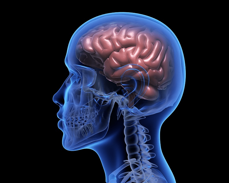
Hemorrhagic strokes make up about 13% of stroke cases. They occur when a weakened vessel ruptures and bleeds into the surrounding brain. The blood accumulates and compresses the surrounding brain tissue.
The two types of hemorrhagic strokes are intracerebral hemorrhage (within the brain) or subarachnoid hemorrhage (between the inner and outer layers of the tissue covering the brain).
What is an arteriovenous malformation?
Normally, arteries carry blood containing oxygen from the heart to the brain, and veins carry blood with less oxygen away from the brain and back to the heart. When an arteriovenous malformation (AVM) occurs, a tangle of blood vessels in the brain bypasses normal brain tissue and directly diverts blood from the arteries to the veins.
How common are brain AVMs?
Brain AVMs are rare, occurring in 10-18 per 100,000 adults. Both sexes are affected equally.
When do AVMs occur?
A small number of AVMs are found at or shortly after birth. But most of them are found later, and likely form and progress later as well.
Where do brain AVMs occur?
Brain AVMs can occur anywhere within the brain or on its covering.
Do brain AVMs change or grow?
Sometimes, AVMs get progressively larger as the amount of blood flow increases.
What are symptoms of a brain AVM?
Symptoms may vary depending on where the AVM is located. This could include a wide variety of brain functions, such as difficulties with movement, coordination, sensation, thinking or memory, speech or vision. Severity can vary greatly.
Symptomatic brain AVMs are found with:
- Hemorrhagic stroke
- Epileptic seizures
- Other symptoms such as progressive neurological deficit
- Headache
What abnormal blood vessels affect the brain?
- True arteriovenous malformation (AVM): The most common brain vascular malformation consists of a tangle of abnormal vessels that connect arteries and veins with no normal intervening brain tissue.
- Cavernous malformations: The vascular malformation in the brain doesn’t actively divert large amounts of blood. It may bleed and often produce seizures.
- Venous malformation: The abnormality is only of the veins.
- Hemangioma: The abnormal blood vessel structures are usually at the surface of the brain and on the skin or facial structures.
- Dural fistula: The covering of the brain is called the “dura mater.” An abnormal connection between blood vessels that involve only this covering is called a dural fistula. It can occur in any part of the brain covering. Three kinds of dural fistulas are:
- Dural carotid-cavernous sinus fistula occurs behind the eye. Patients have eye swelling, decreased vision, redness and congestion of the eye. They often hear a “swishing” noise.
- Transverse-Sigmoid sinus dural fistula occurs behind the ear. Patients usually complain of hearing a continuous noise (bruit) that occurs with each heartbeat, local pain behind the ear, headaches and neck pain.
- Sagittal sinus and scalp dural fistula occurs toward the top of the head. Patients complain of noise (bruit), headaches and pain near the top of the head. They may have prominent blood vessels on the scalp and above the ear.
What causes brain AVMs to bleed?
A brain AVM contains abnormal “weakened” blood vessels that direct blood away from normal brain tissue. These abnormal and weak blood vessels dilate over time. Eventually, they may burst from the high pressure of blood flow from the arteries.
What are the chances of a brain AVM bleeding?
The annual risk of a first-ever ICH from an unruptured brain AVM is ≈1%. The chance of a brain AVM bleeding is about 2% per year over 10 years.
Does one bleed increase the chance of a second bleed?
Yes. The risk of having another episode of AVM bleeding is four times higher than the initial risk.
What can happen if a brain AVM causes a bleed?
Each time blood leaks into the brain, normal brain tissue is damaged. This results in loss of normal function, which may be temporary or permanent. The chance of permanent brain damage is 20% to 30%. The risk of death related to each bleed is 10% to 15%.
How are AVMs diagnosed?
Most AVMs are detected with a computed tomography (CT) brain scan or magnetic resonance imaging (MRI) brain scan. For any type of treatment involving an AVM, an angiogram may be needed to better identify the type of AVM.
What is the best treatment for a dural fistula?
The best treatment is usually endovascular surgical blocking of the abnormal connections that have caused the fistula. This involves inserting small tubes (catheters) inside the blood vessel with X-ray guidance and blocking off the abnormal connections.
What factors influence whether an AVM should be treated?
In general, an AVM may be considered for surgical treatment if it has bled, it’s in an area of the brain that can be easily treated and it’s not too large. The primary goal of treatment is to prevent hemorrhagic stroke.
What is the best treatment for an AVM?
It depends on the type, the symptoms it may be causing and its location and size.
What are different types of treatment?
Medical therapy: If there are no symptoms or almost none, or if an AVM is in an area of the brain that can’t be easily treated, conservative management may be necessary. These patients are advised to avoid excessive exercise and stay away from *blood thinners. (*Some medications are commonly called blood thinners because they can help reduce a blood clot from forming. Common blood thinners are anticoagulants such as warfarin or heparin that slow down the clotting process and antiplatelet drugs such as aspirin and clopidogrel that prevent platelet blood cells from clumping together to build a clot.)
Surgery: If an AVM has bled and/or is in an area that can be easily accessed, then surgery may be recommended. Microsurgery allows the surgeon to work on small structures in the brain using a microscope and small, precise instruments.
Stereotactic radiosurgery: This non-surgical radiation therapy can be used to treat an AVM that’s not too large but is in an area that’s difficult to reach by regular surgery. First, a cerebral angiogram identifies the location of the AVM. Then, high-dose treatment is delivered via targeted radiation on the AVM. This method of radiation therapy causes a scar and allows the AVM to “clot off.”
Interventional neuroradiology/endovascular embolization: It may be possible to treat part or all AVM by placing a catheter inside the blood vessels and blocking off the abnormal vessels with various materials such as glue or coils.
What doctors specialize in treating brain AVMs?
Vascular neurosurgeons specialize in surgical removal.
Radiation therapists/neurosurgeons specialize in stereotactic radiosurgery treatment.
Interventional neuroradiologists/endovascular neurosurgeons specialize in endovascular therapy.
Stroke neurologists specialize in managing brain AVMs.
Neuroradiologists specialize in diagnosing and imaging the head, neck, brain and spinal cord.
What is a cerebral aneurysm?
A cerebral aneurysm is a weak area in a blood vessel that usually enlarges. It’s often described as a “ballooning” of the blood vessel. It could also be called a brain aneurysm or intracranial aneurysm. Cerebral aneurysms can rupture — causing bleeding within the brain or surrounding area.
How do aneurysms form? Are people born with an aneurysm?
People usually aren’t born with aneurysms. They can occur at any age. They’re most common in adults between ages 30 and 60. They occur more often in women than men. Two of the most important modifiable risk factors for cerebral aneurysms are high blood pressure and smoking.
Aneurysms usually develop at branching points of arteries and are caused by constant pressure from blood flow. They often enlarge slowly and become weaker as they grow, just as a balloon becomes weaker as it stretches. Aneurysms may be associated with other types of blood vessel disorders, such as fibromuscular dysplasia, cerebral arteritis or arterial dissection, but these are very unusual. Some aneurysms are due to infections, drugs such as amphetamines and cocaine or direct brain trauma from an accident. About 30,000 ruptured cerebral aneurysms occur each year in the U.S. Up to 6% of the population may have an unruptured cerebral aneurysm.
What are the symptoms of an unruptured aneurysm?
Smaller aneurysms may not have symptoms. As an aneurysm enlarges, it can produce headaches or localized pain. If an aneurysm gets very large, it may produce pressure on the normal brain tissue or adjacent nerves. This pressure can cause difficulty with vision, numbness or weakness of an arm or leg, difficulty with memory or speech, seizures, nausea, vomiting or loss of consciousness.
If one aneurysm forms, will others form?
More than one aneurysm is common; two or more aneurysms are found in 15% to 30% of patients.
What causes an aneurysm to bleed?
Many factors determine whether an aneurysm is likely to bleed. These include the size, shape and location of the aneurysm and symptoms that it causes. Smaller aneurysms that are uniform in size may be less likely to bleed than larger, irregularly shaped ones.
High blood pressure and smoking are risk factors for bleeding. Strenuous activities and strong emotions that cause a sudden and short-lasting rise in blood pressure may lead to a bleeding aneurysm.
*Blood thinners (such as warfarin), some medications and prescription drugs (including diet pills that act as stimulants such as ephedrine and amphetamines), and harmful drugs such as cocaine can cause aneurysms to rupture and bleed.
(*Some medications are commonly called blood thinners because they can help reduce a blood clot from forming. Common blood thinners are anticoagulants such as warfarin or heparin that slow down the clotting process and antiplatelet drugs such as aspirin and clopidogrel that prevent platelet blood cells from clumping together to build a clot.)
What happens if an aneurysm bleeds?
If an aneurysm ruptures, it may leak blood into the space around the brain. This is called a subarachnoid hemorrhage. Depending on the amount of blood, it can produce:
- Sudden severe headache that can last from several hours to days
- Nausea and vomiting drowsiness and/or coma
- Vasospasm, which causes other blood vessels in the brain to narrow and cause limited blood flow
Bleeding can also occur inside the brain. This is called an intracerebral hemorrhage.
Subarachnoid hemorrhage and intracerebral hemorrhage are two types of hemorrhagic stroke. The bleeding damages the brain and can lead to:
- Weakness or paralysis of an arm or leg
- Trouble speaking or understanding language
- Vision problems
- Seizures
- Death
What is the usual damage to the brain after an aneurysm bleeds?
Once an aneurysm has bled, re-bleeding is a risk.
About 25% of people whose cerebral aneurysm has ruptured don’t survive the first 24 hours. If the aneurysm isn’t treated quickly enough, another bleed may occur from the already ruptured aneurysm.
Vasospasm (irritation by the leaked blood causing narrowing of the blood vessels) is a common complication following a ruptured aneurysm. This can lead to further brain damage.
Other problems may include hydrocephalus (enlargement of the spaces within the brain that produce cerebrospinal fluid), difficulty breathing that requires a mechanical ventilator and infection.
Why is the damage so extensive after bleeding?
After blood enters the brain and the space around it, direct damage to the brain tissue and brain function results. The amount of damage is usually related to the amount of blood. Damage is due to the increased pressure and swelling from bleeding directly into the brain tissue, or from local cellular damage to brain tissue from irritation of blood in the space between the brain and the skull.
Blood can also irritate and damage the normal blood vessels and cause vasospasm (constriction). This can interrupt normal blood flow to the healthy brain tissue and can cause even more brain damage. This is called an ischemic stroke.
How is an aneurysm diagnosed?
Special imaging tests can detect a brain aneurysm. In the CTA (computed tomographic angiography), patients are placed on a table that slides into a CT scanner. A special contrast material (dye) is injected into a vein, and images are taken of the blood vessels to look for abnormalities such as an aneurysm. In the second test, called MRA (magnetic resonance angiography), patients are placed on a table that slides into a magnetic resonance scanner, and the blood vessels are imaged to detect a cerebral aneurysm.
Cerebral angiogram is most reliable in identifying the exact location, size and shape of aneurysms, and can be useful to fully map a plan for therapy. In this test, the patient lies on an X-ray table. A small tube (catheter) is inserted through a blood vessel, usually in the leg (groin), and guided into each of the blood vessels in the neck that go to the brain. Contrast is then injected, and pictures are taken of all the blood vessels in the brain. This test is slightly more invasive and less comfortable.
To develop a full picture to diagnose the aneurysm and to determine the best treatment, other tests could include a CT scan, MRI, MRA and cerebrospinal fluid (CSF) analysis.
What follow-up is required after aneurysm treatment?
Depending on the type of treatment, the two follow-up procedures are:
- Surgical clipping: After this type of surgery, a post-operative angiogram is usually performed during the hospital stay to make sure the surgical clip has completely treated the aneurysm.
- Angiogram: After coiling an aneurysm, a routine follow-up angiogram is usually performed six to 12 months after the procedure to make sure the aneurysm remains blocked off.
Will treating a ruptured aneurysm reverse or improve brain damage?
Once an aneurysm bleeds and the brain is damaged, treating the aneurysm won’t reverse the damage. Treatment helps prevent more bleeding.
How do doctors choose a treatment method for an aneurysm?
Doctors evaluate the risk factors that favor treatment versus non-treatment and decide which technique may be best. It’s important to consult with experts in this field. This should include a cerebrovascular neurosurgeon who specializes in surgically clipping aneurysms, a neurosurgeon with endovascular expertise and training, a neurointerventionalist (a neurologist with endovascular training) or a neuroradiologist who specializes in the less-invasive treatment of cerebral aneurysms by coiling.
How should an aneurysm be treated?
The treatment time and options depend on the size, location and shape of the aneurysm, symptoms and the patient’s overall medical condition. A ruptured aneurysm usually requires treatment right away.
Small, unruptured aneurysms that aren’t creating symptoms may not need surgical treatment unless they grow, trigger symptoms or rupture. It’s important to have annual checkups to monitor blood pressure, cholesterol and other medical conditions.
Neurosurgery
Depending on a person’s risk factors, open surgery may be recommended. Patients are placed under general anesthesia, and the neurosurgeon places a surgical clip around the base of the aneurysm. After this type of surgery, a post-operative angiogram is usually performed during the hospital stay to make sure the surgical clip has completely treated the aneurysm.
Neurointervention
Depending on the aneurysm’s size, location and shape, it may be treatable from inside the blood vessel. This minimally invasive procedure is similar to the cerebral angiogram. However, in addition to taking pictures, a catheter is directed through the blood vessels into the aneurysm. Then, using X-ray guidance, the endovascular surgeon carefully places soft platinum micro-coils into the aneurysm and detaches them. The coils stay within the aneurysm and act as a mechanical barrier to blood flow, thus sealing it off. After coiling an aneurysm, a routine follow-up angiogram is usually performed six to 12 months after the procedure to make sure the aneurysm remains blocked off.
What are the potential complications of aneurysm treatment?
Until the aneurysm is safely and completely treated, re-bleeding and more brain damage is a risk. If normal blood vessels are damaged, it could also result in more brain damage.

Read Eileen’s storySurvivor Story: Eileen Haas
Eileen Haas was folding laundry when she felt something “break” in her head. There was the sensation that something warm was cascading down her back, as if a blood vessel had burst. Lying on the floor in her second-floor bedroom, unable to move, Haas heard ambulance sirens growing closer and then the sound of paramedics running up the stairs.
Doctors told her that she had suffered a hemorrhagic stroke, or bleed in her brain.
